Horse Hoof Anatomy Explained: What Every Part of the Hoof Does
The hoof looks like a simple block of horn, but under it sits a bone, a joint, a tendon and hundreds of interlocking layers holding a whole horse up. Know what each part does and every time you pick out a foot, you’re reading it.
I learned that lesson at sixteen, with my very first horse. Before he came to me he had fallen off a truck and sliced away the whole inside heel bulb on his off fore. By the time he was mine it had healed, but that foot was never quite like the other three. It needed checking every single day, and I learned what was normal for it and what wasn’t. It’s an early lesson I’ve never forgotten: you can’t look after a horse’s feet properly until you understand what you’re looking at.
Free Hoof Health Toolkit
A weekly hoof check sheet for all four feet, a “when to call the vet” chart and a farrier & vet record. Print it and pin it up.
Download Free →The Outside: Hoof Wall and Coronary Band
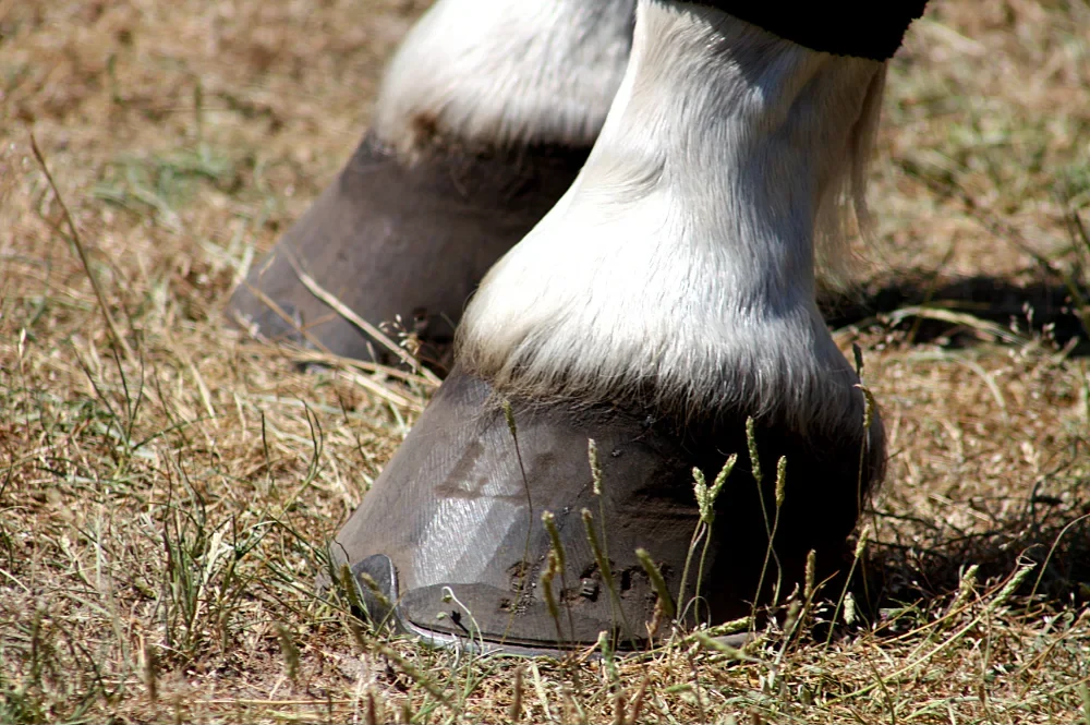
Everything you can see when the horse is standing is the hoof wall. It’s made of keratin, the same protein as your fingernails, laid down in tiny tubes that run from the top of the hoof to the ground. Those tubes give the wall its strength, a bit like the grain in a plank of wood.
Toe, quarters and heels
The wall is thickest and steepest at the toe, thins out round the quarters (the sides), and is lowest and most flexible at the heels. That flexibility matters: the back of the foot is meant to spread slightly every time it takes weight.
The coronary band
Running round the top of the hoof where the hair stops is the coronary band (coronet). This is where almost all of the wall is grown, so anything that injures or stresses the coronet shows up later as a ridge, a crack or a weak patch growing down the wall. Just below it is a thin, shiny layer called the periople, which works like the cuticle on your nail.
Horizontal rings on the wall are a record of what happened at the coronet months ago. Faint, even rings are usually just changes in diet or season. Rings that are wider at the heel than at the toe can be a sign of a past laminitis episode and are worth showing your vet or farrier.
Underneath: Sole, Frog, White Line and Bars
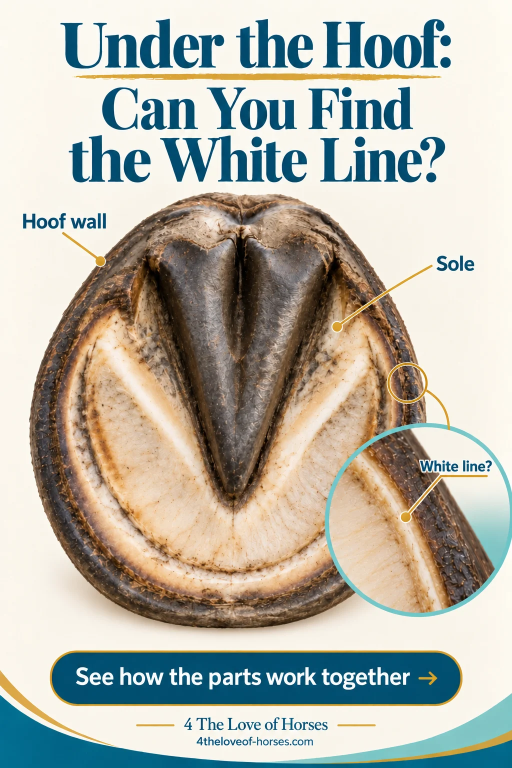
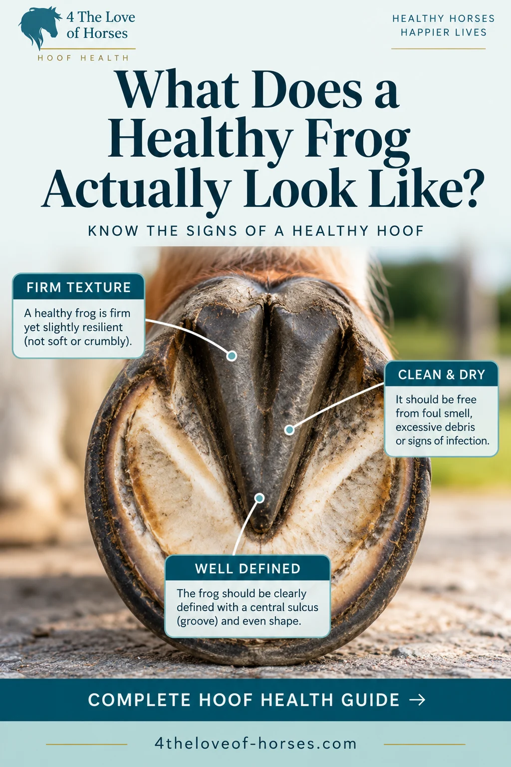
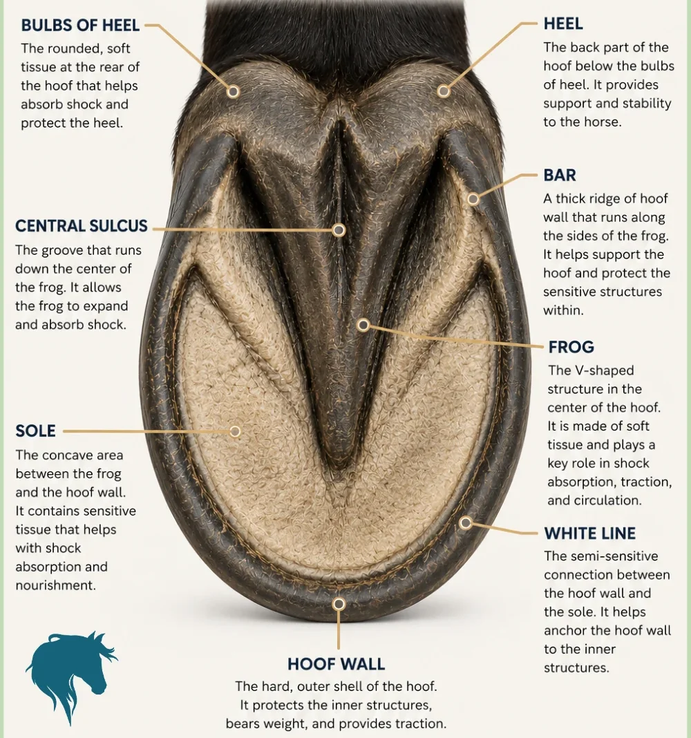
Pick up a front foot and this is roughly what you’re looking at. It’s the view you get every day, and the one where most problems first show themselves.
Sole
Protects the inside of the foot from the ground. It should be slightly dished (concave) so it doesn’t take the horse’s full weight. Old sole flakes away naturally; that chalky shedding is normal.
Frog
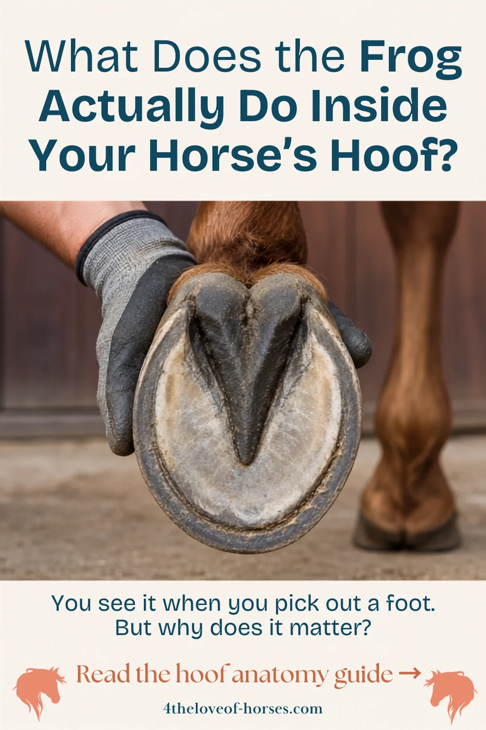
The rubbery V-shape in the middle. It grips, absorbs shock and helps the heels spread on landing. A healthy frog feels firm and springy, like a pencil eraser.
White line
The narrow, pale band where the wall joins the sole. It’s softer than the wall around it, which is why stones, gravel and infection tend to get in here first.
Bars
The wall folds forward at each heel to form the bars, which brace the back of the foot and stop the heels collapsing inwards.
Grooves (sulci)
The grooves either side of the frog and the dip down its centre. They should be shallow and clean. A deep, narrow central groove is a favourite hiding place for thrush.
Heel bulbs
The soft, rounded back of the foot. They sit over the digital cushion and take the first contact when a horse lands heel-first, as it should.
The angle between the wall and the bar at each heel is the seat of corn. It’s where corns (bruises) appear, often from a shoe left on too long so that the heel of the shoe ends up pressing on the sole. Reddish or purple staining here at a trim is worth a question to your farrier.
Inside the Hoof: Bones, Laminae and the Digital Cushion
The hoof capsule is a protective case. Inside it is living, sensitive tissue with a rich blood supply, which is why problems inside the foot are so painful.
The bones
- Coffin bone (pedal bone, P3): the main bone inside the hoof, shaped roughly like a small hoof itself. It carries the horse’s weight and is the bone that rotates or sinks in severe laminitis.
- Short pastern bone (P2): sits partly inside the top of the hoof and meets the coffin bone at the coffin joint.
- Navicular bone: a small, boat-shaped bone behind the coffin joint. The deep digital flexor tendon runs over it like a rope over a pulley, which is why the back of the foot is under so much pressure, and why navicular syndrome develops there.
The laminae: what holds the bone in place
The coffin bone isn’t resting on the sole; it’s hanging from the inside of the hoof wall. Around 550 to 600 leaf-like folds called laminae on the bone interlock with matching folds on the inside of the wall, and each of those has a hundred or more smaller folds of its own. Think of two hairbrushes pushed together, bristles meshed, with the whole horse’s weight on the join.
When those laminae become inflamed and weaken, that’s laminitis. If enough of the bond fails, the coffin bone can pull away from the wall (rotate) or sink down towards the sole. This is why laminitis is always an emergency, and why the white line, the only part of this bond you can see from underneath, is worth looking at every day.

The digital cushion and collateral cartilages
Filling the back half of the foot, above the frog, is the digital cushion, a wedge of tough, springy tissue that soaks up concussion. Either side of it are the collateral cartilages, two flexible plates you can feel just above the coronet at the heels. In some older horses, especially heavier types, these cartilages slowly turn to bone. That’s called sidebone, and most horses with it are never lame from it.
The digital cushion isn’t fixed at birth. It gets thicker and firmer in horses that move a lot on varied ground, and stays thin and soft in horses that live in stables on deep bedding. It’s one of the clearest examples of the foot adapting to how the horse lives.
How It All Works Together
Every time the hoof lands, the heels and frog take the first contact. The back of the hoof spreads slightly, the digital cushion compresses and the cartilages flex outwards. As the foot lifts, everything springs back. This is often called the hoof mechanism.
That spring-and-release does two jobs. It takes the sting out of each stride before it travels up the leg, and it helps move blood through the foot’s dense network of vessels. Exactly how much the frog acts as a “pump” is still debated, but there’s no argument that horses which move more have better circulation in their feet, and that feet which can’t flex properly (contracted heels, a frog that never touches the ground) don’t function as well.
Movement is good for feet. Turnout, walking on varied ground and regular trims that keep the heels and frog doing their job all feed straight back into how the hoof is built. A horse standing in a stable 20 hours a day is missing most of that.
Balance and the hoof-pastern axis
Look at your horse’s foot from the side while he stands square. A straight line should run down the front of the pastern and carry on down the front of the hoof wall. When the toe is long and the heels low, that line “breaks back”; when the heels are too high, it “breaks forward”. Either way, more strain lands on certain structures, usually the tendons, the navicular area or the laminae at the toe. Your farrier is trimming to that line, not to a fixed angle.
Which Part Goes Wrong, and What It Looks Like
Once you know the parts, the common problems make a lot more sense. Most of them start in one structure.
| Structure | Common problem | Early sign you can spot |
|---|---|---|
| Frog grooves | Thrush | Black, smelly material in the grooves; ragged frog |
| White line | White line disease, gravel, abscesses | Dark spots, widening or crumbly material where wall meets sole |
| Laminae | Laminitis | Strong digital pulses, heat, rocked-back stance, reluctance to turn |
| Hoof wall | Cracks, flares, chips | New cracks, especially from the coronet; wall flaring out at the quarters |
| Sole | Bruising, thin soles | Sore on stony ground; red or purple marks at a trim |
| Seat of corn | Corns | Lameness after a shoe has been on too long; staining at the heel |
| Navicular area | Navicular syndrome | Short, choppy steps; landing toe-first; pointing a front foot |
| Collateral cartilages | Sidebone | Hard, bony feel just above the coronet at the heels |
Sudden severe lameness, strong pulses with heat in both front feet, or anything stuck in the sole needs your vet the same day. Leave a nail or sharp object in place if you safely can, as the vet may want to X-ray it to see which structures it has reached. Our signs your horse needs the vet right now guide covers the rest.
Put this into practice every day
The Hoof Health Toolkit turns this into a tick-sheet for all four feet, plus how to feel a digital pulse and a red, amber, green “when to call” chart.
Get the Free Toolkit →Questions Owners Ask
Does a horse feel anything in its hoof?
The outer wall and the outer layers of the sole have no nerves, which is why a farrier can trim and nail into the wall without hurting the horse. Everything inside, including the laminae, the coffin bone, the sole’s living layer and the digital cushion, is very sensitive. A nail placed too far in, a bruise or an abscess is genuinely painful.
Should the frog touch the ground?
On a horse standing on soft ground, yes, the frog should be close to level with the wall at the heels so it shares the load. On hard ground it may sit a little higher. A frog that never makes contact tends to shrink and the heels can contract. Your farrier will usually leave a healthy frog alone and only trim away loose, ragged tags.
How long does it take to grow a new hoof?
About 9 to 12 months for the toe and 4 to 6 months at the heels. So if you change your horse’s feed or trimming, you’re judging the results on growth that’s still months from the ground. Patience is part of hoof care.
What’s the difference between the coffin bone and the pedal bone?
Nothing. They’re two names for the same bone, also called P3 or the third phalanx. You’ll hear “pedal bone” more in the UK and South Africa and “coffin bone” more in the US.
Are front and hind hooves the same?
No. Front feet carry around 60% of the horse’s weight and are rounder and wider. Hind feet are narrower, more pointed at the toe and a little steeper. Comparing pairs (front with front, hind with hind) is how you spot a foot that’s changing shape.
Keep Learning
Further reading: Foot Anatomy, Large Animal Surgery (University of Minnesota open textbook) · Horse Hoof Anatomy, The Horse. This article is general information; always involve your vet and farrier for diagnosis and treatment.